Our retinal projects are organised around several major themes:
Funded by the Macular Society, we have been able to create a disease model in the lab which we can use for various studies
Age related macular degeneration (AMD) is the commonest cause of blindness in the developed world. There are two manifestations of advanced AMD resulting in loss of vision: dry and wet; the dry type being the most common and with no treatment available. Understanding what happens in AMD affected retina is a crucial basis for developing therapies. Funded by the Macular Society, our lab generated a cell model of AMD by turning skin cells from people affected by AMD into retinal pigment epithelium (RPE) cells, the principle cell type that gets damaged and lost during the course of AMD (Hallam et al. 2017, Stem Cells. 2017 Nov;35(11):2305-2320). Our work has shown that AMD-RPE cells have expanded and less functional lysosomal compartments, which are organelles that are involved in digestion of unwanted intracellular materials (autophagy) (Cerniauskas et al. 2020, Stem Cells Transl Med. 2020 Dec;9(12):1585-1603). We found that lysosomal membranes are fragile and this may cause the leakage of their crucial digestive enzymes and their deposition into drusen. Drusen are build-ups of waste products under the RPE and large accumulations of drusen are the defining feature of macular degeneration. We have also shown that the probable cause of the lysosomal deficiency is uncontrolled activation of the complement system, which is part of the immune system that clears pathogens. Treatment with a specific complement inhibitor that stops this activity helped to stop the progression of AMD features in AMD-RPE cells, which are promising results suggesting new treatment options for AMD
Our further work, funded by the Macular Society, has shown that AMD-RPE cells produce stress markers that are moved outside the cells in tiny vesicles called exosomes, which help cells to communicate with each other (Kurzawa-Akanbi at al. J Extracell Vesicles, 2022 Dec;11: e12295). We are able to isolate these vesicles and analyse their contents. We found that AMD-RPE produce around twice as many exosomes compared to RPE cells derived from people without AMD. AMD-RPE exosomes are very harmful to healthy RPE cells and make them develop AMD-like features, which makes us believe they are involved in the progression of AMD. We are now in a process of careful examination of AMD-RPE exosomes contents, which will allow us to pinpoint specific exosomal markers that could potentially be used to detect AMD. As exosomes are tiny, they can enter the bloodstream and we are hoping to be able to detect those AMD-RPE stress related exosomal markers in the patients’ blood, which could give us means to spot AMD early and predict the progression of the disease (dry or wet type).
As our studies implicate faulty autophagy in macular degeneration, we are looking into other macular diseases that may be due to similar cellular defects. Mutations in DRAM2, which is an autophagy regulator, are known to cause retinal degeneration with early macular cone photoreceptor involvement. Our lab generated induced pluripotent stem cell lines (iPSC) from patient skin cells with DRAM2 mutations. We have been able to erase these mutations using the CRISPR technology (CRISPR) and produce controls for the disease mutation-bearing retinal cells. Using these stem cells, we generated retinal organoids (“retinas in a dish”) models and RPE monolayers that allowed us to identify a key role for DRAM2 in maintaining the integrity of photoreceptors and RPE cells by regulating lysosomal function, autophagy, and potentially vesicular trafficking (Tsikandelova et al, Stem Cell Reports 2024 Aug 13;19:1107-1121).
Growing evidence indicates that damage to choriocapillaris endothelial cells (CECs), the principal components of the tiny blood vessels beneath the retina in the choroid, plays a crucial role in AMD development and progression. These vessels supply the retina with oxygen and nutrients while removing metabolic waste, thereby supporting the high energy demands of the retinal pigment epithelium (RPE) and photoreceptors essential for healthy vision. Studies of human donor eyes reveal that CECs undergo early degeneration in AMD, leaving behind characteristic “ghost” vessels. Our recent single-cell transcriptomic analyses reinforce these observations, identifying CECs beneath the macula as particularly vulnerable. This suggests that strategies to preserve or restore CEC integrity could slow or prevent AMD progression. In our ongoing project, we generate CECs from PSCs derived from AMD patients and employ CRISPR/Cas9 gene editing to produce genetically corrected controls. By comparing these cell types, we aim to uncover mechanisms underlying AMD-associated CEC loss and to identify compounds capable of protecting them. Such interventions could enable earlier therapeutic action, preserving central vision and helping patients maintain independence and quality of life.
Transforming ophthalmic research
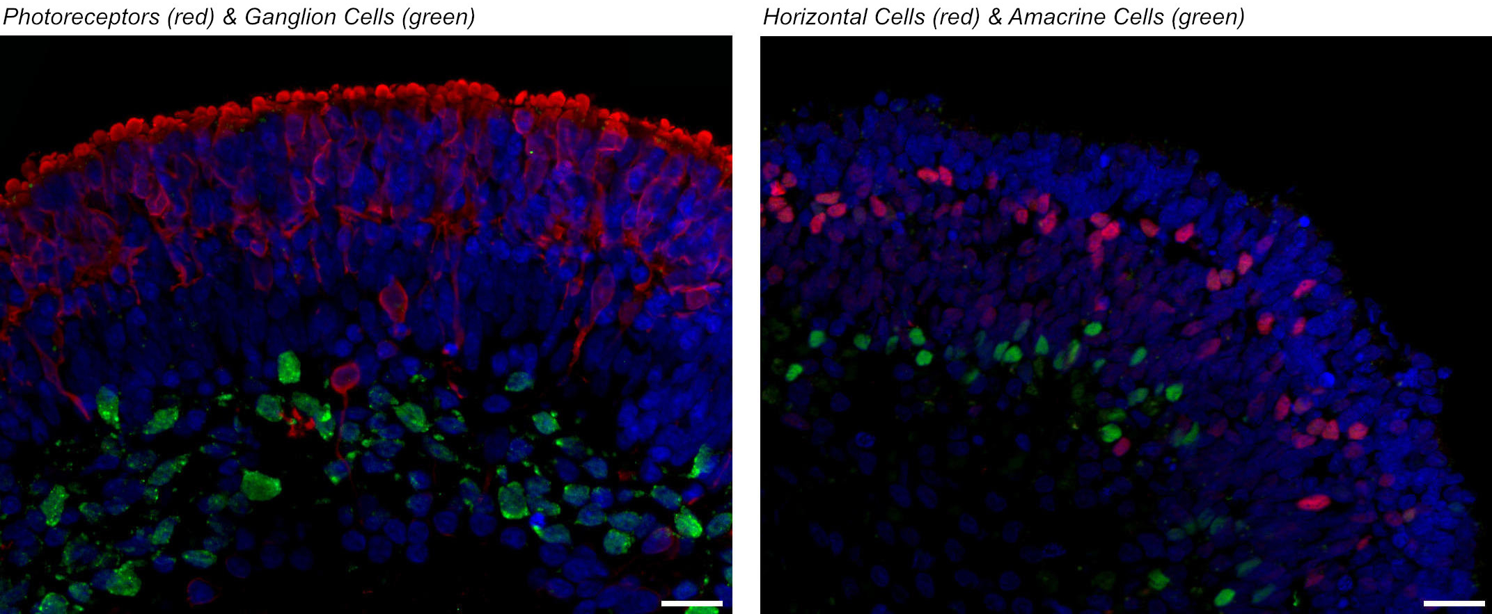
The generation of human retinal tissue from stem cells under laboratory conditions has been a pioneering breakthrough which has transformed the field of ophthalmic research. In the last five years, our group has developed robust methods for the large-scale generation of retinal organoids (retina in a dish) from human embryonic and human induced pluripotent stem cells. These mini retinas in a dish contain all retinal cell types and display the same five layered structure as known from adult retina. Moreover, they exhibit the most crucial function of the retina, light sensitivity. Our group is using these mini retinas for:
Retinal organoids derived from human pluripotent stem cells.
These projects have benefited from multiple funding awards (European Research Council, Retina UK and NCR3Rs) and close collaborations with Prof. Evelyne Sernagor at the Bioscience Institute (Newcastle University) and Dr Gerrit Hilgen (Northumbria University).
iPSC Models of PRPF31/8 Retinitis Pigmentosa: Spliceosomal Defects and Therapeutic Insights
Retinitis pigmentosa (RP) is the most common inherited retinal disease characterized by progressive degeneration of photoreceptors leading to night vision loss, restricted visual field until eventual blindness. The prevalence of RP is around 1 in 4000 and there are over 1.5 million RP patients worldwide. Mutations in the pre-mRNA processing factor (PRPF) genes encoding the core spliceosome components account for 15-20% of autosomal dominant RP cases. Using reprogramming approaches and CRISPR/Cas9 technologies our group has generated patient specific iPSC lines from patients with PRPF31 and PRPF8 mutations. Our work has shown severe RPE defects resulting from PRPF31 mutations, including global spliceosome dysregulation, disrupted apical-basal polarity, reduced trans-epithelial resistance, compromised phagocytic capacity, decreased cilia length and incidence. Disrupted cilia morphology also occurred in patient-derived photoreceptors, associated with progressive degeneration and cellular stress (Buskin et al. Nat Commun. 2018 Oct 12;9(1):4234).
Quantitative proteomics and functional analyses revealed progressive accumulation of cytoplasmic protein aggregates in PRPF31-mutant RPE, affecting RNA splicing, autophagy–lysosome and unfolded protein response pathways, and demonstrated that pharmacological activation of autophagy can clear these aggregates and restore key RPE functions, highlighting a tractable therapeutic strategy for PRPF31-RP (Georgiou et al. Clin Transl Med. 2022;12:e759).
Funded by the MRC and in collaboration with research groups led by Prof. Colin Johnson (University of Leeds), Prof. Robin Ali (King’s College, London) Dr. Sina Mozaffari-Jovin (Max Planck Institute, Gottingen) and Dr Sushma Nagaraja-Grellscheid (University of Bergen), we also investigated the impact of PRPF8 mutations on retinal cells. Similarly, PRPF8 mutations cause retinal degeneration through spliceosome dysfunction; patient-derived RPE and retinal organoids exhibited splicing defects, photoreceptor loss, polarity disruption, ciliary abnormalities, cryptic splice site activation, and proteome changes linked to degeneration pathways, underscoring shared mechanisms across PRPF mutations (Atkinson et al. Nat Commun. 2024; 15:3138).
The potential to investigate pathological changes in the human eye
Retinoblastoma is a childhood cancer of the developing retina, the light-detecting tissue of the eye. Biallelic mutations in RB1 gene encoding pRB (retinoblastoma protein) accounts for ~98 % of retinoblastoma cases in young children (up to five years old).
The palette of pRB degenerative mutations that define malignant signature and the demand to establish enhanced clinical protocols of treatment that not only ensures child’s survival but also normal development post-treatment hence low toxicity with high efficacy, cannot make a more compelling case for further studies.
Well established in our laboratory methodology for retinal organoids formation and the enormous potential of this 3D model to investigate pathological changes in the human eye encouraged us to establish in vitro model of retinoblastoma, initiated by biallelic inactivation of RB1. This project benefited from Fight from Sight funding and close collaborations with Consultant Ophthalmologist, Mr. Manoj Parulekar.
To obtain the model that resembles patient-specific genomic background in combination with a hereditary mutation in the RB1 gene, we generated iPSC lines from two patients with RB1 mutations. Retinal organoids made from patient iPSCs lacking expression of the RB1 gene displayed the hallmarks of retinoblastoma tumours and responded predictably to key chemotherapeutic substances used for treatment of the disease (Rozanska et al. Stem Cells Transl Med. 22022; 11:415-433). Funded by the Little Princess Trust, we have undertaken screening of 39 compounds, with 2 promising leads to take forward in pharmacogenetic studies.
There is a need for research into replacement of photoreceptors
Our retinas exhibit limited regenerative capacity; hence, there is a need for research into replacement of photoreceptors in patients with retinal disease. Our aim is to study and improve photoreceptors transplantation with the aim of developing therapeutic interventions to restore vision at advanced stages of retinal degeneration.
With the advances made in differentiation of human pluripotent stem cells (hPSCs), we are able to generate physiological functional retinal organoids which contain photoreceptors alongside other retinal neurons and Müller glial cells arranged in a laminated structure resembling the native retina. We have developed pre-clinical tools for enrichment of photoreceptors (Collin et al., 2016; Stem Cells. 2016; 34:311-21), which we deliver as cell suspensions into the subretinal space (Collin et al. 2019; Stem Cells. 2019; 37:609-622). In close collaboration with Prof. Evelyne Sernagor (Newcastle University) and Dr. Gerrit Hilgen (Northumbria University), we demonstrated robust donor–host integration and evidence of synaptic connectivity within degenerate host retinas. Transplanted cone photoreceptors exhibited mature outer segment morphology and expressed key visual transduction proteins, correlating with significant restoration of visual responses at both retinal and behavioural levels. These findings validate the functional potential of organoid-derived photoreceptors for retinal repair and provide a strong basis for further optimisation of cell delivery and integration strategies (Zerti et al. Stem Cells. 2021; 39:882-896). Funded by the MRC, we are assessing the most optimal rod-cone transplant in animal models of retinal degeneration for the treatment of advanced Retinitis Pigmentosa.
![]()
Promising treatment option for inherited blindness
Gene therapy is a medical approach where engineered genetic material is introduced into human cells to treat hereditary diseases. Gene therapy corrects pathogenic variants of diseases by replacing the defective gene with a functional copy, silencing of the mutated gene, or by inserting or overexpressing a corrected version of the mutated gene. The corrected genes will then be transcribed using the host’s transcription machinery to produce functional proteins and reverse disease phenotypes.
Over the past decade, recombinant AAV (rAAV) has emerged as a useful delivery vector for the in vivo delivery of retinal gene therapies, and the first approved target for ophthalmological treatment. Most ongoing clinical trials for inherited retinal disorders are AAV-mediated gene therapies. The therapeutic effects of AAV-mediated gene therapy was first studied on Leber Congenital Amaurosis caused by a pathogenic mutation in the RPE65 gene (RPE65-LCA). The success of LUXTURNATM (voretigene neparvovec-ryzl; Spark Therapeutics Incorporate, USA) as the first FDA-approved gene therapy for treating biallelic RPE65-retinal dystrophy (NTC00999609) was a huge milestone and accelerated gene therapy research (US Food & Drug Administration, 2017).
In our recent collaboration with Prof. Robin Ali’s ocular gene therapy research group (King’s College London), we are assessing the safety and efficacy of AAV-GT in ameliorating pathological phenotypes of several forms of inherited retinal disorders, including Retinitis Pigmentosa (RP) and Cone-rod Dystrophy 21 (CORD21). By targeting the mutated PRPF31 and DRAM2 genes respectively. This promising treatment option is assessed using biologically relevant in vivo disease models, including 2D retinal pigmented epithelial culture (RPE) and 3D retinal organoids (ROs) derived from patient-specific induced pluripotent stem cells.
We are actively participating in current clinical trials
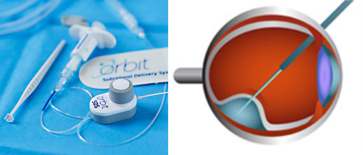
Our group is also actively participating in clinical trials of advanced therapies and hope to develop our own studies. Professor Steel participated as a Phase 1 surgeon in the ground-breaking Astellas trial of embryonic stem cell-derived RPE cell transplantation into the subretinal space in patients with Stargardt's macular dystrophy and is now in set up to evaluate this treatment in patients with early AMD. He was the one of the first UK surgeons to deliver subretinal gene therapy for dry AMD as part of the phase 1 FOCUS study with Gyroscope Therapeutics and is similarly soon to start the Complement therapeutics phase 1 study subretinal gene therapy study evaluating a novel complement regulator. Notably, he is one of only a few global surgeons trained in a novel suprachoroidal subretinal delivery technique, soon to be used in several global studies and anticipated to offer several advantages to conventional trans-vitreal delivery.
As well as these advanced therapeutics, he has also acted as both principal and chief investigator for over 50 clinical trials and is currently leading or assisting several UK multicentre trials evaluating novel and improved treatments. These include the use of air in rhegmatogenous retinal detachment surgery, the treatment of submacular haemorrhage, gasless macular hole surgery, the timing of surgery for severe diabetic eyes disease, and the use of combined lens surgery in RRD. He will also be the first non-US principal investigator for the world-leading DRCR network, the largest retinal research network globally, conducting a study on the timing of surgery for epiretinal membranes. Finally, he is also leading a major regional health initiative and new eye research institute as Co-director of Northern Ophthalmic Research and Innovation (NORI) centre themed on improving population health using eye imaging-based oculomics.
Our aim is to understand how and when parts of our eyes form, function and interact
Vision loss can occur through the effect of faulty genes we inherit from our parents as well as the accumulation of damage and the effect of various diseases throughout our lives.
Our ability to prevent and treat vision loss is closely linked with our knowledge of how eyes form, how they work and when and what is likely to go wrong. The availability of tissue to study during pregnancy and through adulthood is very limited. Our group is in a unique position, having access to eyes through resources such as the HDBR, a collaboration between our University and University College London, which collects samples from aborted embryos and foetuses with the mother's consent, and NHSBT.
Our aim is to use these samples to understand how and when parts of our eyes form, function and interact, and look at the role of genes that cause loss of vision when faulty. This work commenced in 2011 and we have compiled images of and information on genes involved in the developing and adult eye, which we have reported in Mellough et al Development. 2019 Jan 29;146(2): dev169474). Funded by BBSRC, we have completed this work at the single cell level, generating new insights into the location of retinal progenitor cells during early human development (Dorgau et al. Nat Commun. 2024; 15:3567, Dorgau et al. iScience. 2024; 27:109397).
Retinal Stem Cell Research
Biosciences Institute
Newcastle University, International Centre for Life, Central Parkway, Newcastle upon Tyne, NE1 3 BZ. United Kingdom
Tel: +44 (0)191 241 8688
Email:
majlinda.lako@ncl.ac.uk